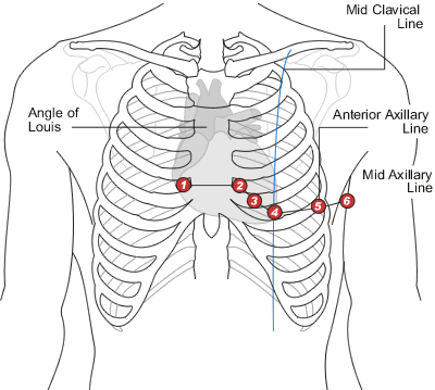
Diagram Of Chest Area : Coronary Artery Bypass Graft Cabg Nhs - The ribs and sternum make up what is called the 'ribcage.' the ribcage protects the lungs, blood vessels, and heart.
Diagram Of Chest Area : Coronary Artery Bypass Graft Cabg Nhs - The ribs and sternum make up what is called the 'ribcage.' the ribcage protects the lungs, blood vessels, and heart.. The ribs and sternum make up what is called the 'ribcage.' the ribcage protects the lungs, blood vessels, and heart. Diagram of the emergence of the bronchial arteries in the descending thoracic aorta. Chest pain or discomfort that is new, worsening, or occurs at rest. It is a flat bone about six inches in length, around an inch wide, and only a fraction of an. Nerves provide sensation to the breast.
The pectoralis major originates along the clavicle, down the sternum, and across the ribs and inserts into the humerus. The aorta is the large artery leaving the heart. It lies between the right and left lungs, in the middle of the chest and slightly towards the left of the breastbone. Chest pain can also be due to a heart attack (coronary occlusion), aortic aneurysm dissection, myocarditis, esophageal spasm, esophagitis. Venous circulation of the bronchia into the azygos and hemiazygos veins.

It also protects several vital organs of the chest, such as the heart, aorta, vena cava, and thymus gland that are located just deep to the sternum.
These areas are specialized for. Related posts of anatomy of the chest area pressure points on female anatomy. The throat is one of the most complex parts of the human body. A person with chest pain on the left side may be experiencing lung problems. Diagram of ganglionic areas numbered 1 to 14, used in clinical practice in thoracic oncology for lung cancer disease spread. There are many causes of chest pain.a serious form of chest pain is angina, which is a symptom of heart disease and results from inadequate oxygen supply to the heart muscle. The diaphragm, a sheet of muscle in the middle chest area, is essential for breathing. A person with a muscle strain in the chest may experience sudden, sharp pain in this area. The myofascial pain pattern has pain locations that are displayed in red and associated trigger points shown as xs. The circulatory system does most of its. The sternum, or breastbone, is a flat bone at the front center of the chest. However, there are three indications. Nerves provide sensation to the breast.
Understanding the basics of throat anatomy with diagram and pictures. A typical heart is approximately the size of your fist: Location of chest pain during angina or heart attack diagram in this image, you will find an upper chest, substernal radiating to neck and jaw, substernal raiding down left arm, substernal radiating down left arm, epigastric radiating to neck, jaw, and arms, neck and jaw, left shoulder and down both arms, intrascapular in it. You also may feel an ache in your back or right shoulder blade. The shape of the heart is similar to a pinecone, rather broad at the superior surface and tapering to the apex.

The sternum is located along the body's midline in the anterior thoracic region just deep to the skin.
In the lungs, the pulmonary arteries (in blue) carry unoxygenated blood from the heart into the lungs. It can be difficult to identify whether chest pain is a sign of a heart attack. Chest pain or discomfort that is new, worsening, or occurs at rest. These areas are specialized for. The epidermis is the outermost layer that provides a protective, waterproof seal over the body. Pressure points on female anatomy 11 photos of the pressure points on female anatomy female dog names, female pleasure points, female pressure points diagram, male pressure points, pressure points on female body, human anatomy, female dog names, female pleasure points, female pressure points diagram, male. Thoracic cavity, also called chest cavity, the second largest hollow space of the body.it is enclosed by the ribs, the vertebral column, and the sternum, or breastbone, and is separated from the abdominal cavity (the body's largest hollow space) by a muscular and membranous partition, the diaphragm.it contains the lungs, the middle and lower airways—the tracheobronchial tree—the heart. The nervous system of the thorax is a vital part of the nervous system as a whole, as it includes the spinal cord, peripheral nerves, and autonomic ganglia that communicate with and control many vital organs. It lies between the right and left lungs, in the middle of the chest and slightly towards the left of the breastbone. Sensory information from the body and critical signals. A person with a muscle strain in the chest may experience sudden, sharp pain in this area. Any diaphragm pain can, therefore, be very alarming. When your gallbladder gets inflamed and swollen, symptoms include pain in your belly, including the area just above your stomach.
The major muscle in the chest is the pectoralis major. Pectoralis major trigger point diagram, pain patterns and related medical symptoms. Angina can be caused by coronary artery disease or spasm of the coronary arteries. This is an emergency situation as it can precede a heart attack, serious abnormal heart rhythm, or. Possible causes of pain include trauma, musculoskeletal.

The superior vena cava is the large.
Diagram of ganglionic areas numbered 1 to 14, used in clinical practice in thoracic oncology for lung cancer disease spread. When your gallbladder gets inflamed and swollen, symptoms include pain in your belly, including the area just above your stomach. The myofascial pain pattern has pain locations that are displayed in red and associated trigger points shown as xs. Shape and size of the heart. Diagram of the emergence of the bronchial arteries in the descending thoracic aorta. A typical heart is approximately the size of your fist: The aorta is the large artery leaving the heart. This pericardium is attached to the diaphragm, spinal column and other parts via strong ligaments. Chest pain or discomfort that is new, worsening, or occurs at rest. A woman's chest — like the rest of her body — is covered with skin that has two layers. This is an emergency situation as it can precede a heart attack, serious abnormal heart rhythm, or. The terms pulled muscle and muscle strain refer to an injury that involves an overstretched or torn muscle. The chest is the area of origin for many of the body's systems as it houses organs such as the heart, esophagus, trachea, lungs, and thoracic diaphragm.
Komentar
Posting Komentar